

Viruses are often termed “the invisible enemy”. They aren’t visible with the naked eye, or even by using a standard optical microscope. So how do we know they exist or what they look like?
There are biochemical methods, such as the ones used to confirm COVID-19 infection, that look for evidence of genetic material from a virus. But there are also multiple different methods we use in the laboratory to “see” viruses.
To understand these methods, we first need to understand how small viruses actually are. Most of our cells are around 100 micrometres (0.1 millimetres) in diameter. Viruses are about 1,000 times smaller than this averaging around 150 nanometres (0.00015 millimetres).
Light microscopy


Standard light microscopes allow us to see our cells clearly. However, these microscopes are limited by light itself as they cannot show anything smaller than half the wavelength of visible light – and viruses are much smaller than this.
But we can use microscopes to see the damage viruses do to our cells. We call this “cytopathic effect”, and comparing infected cells to uninfected ones enables us to detect the presence of viruses in a sample.
Preliminary work on SARS-CoV-2 (the virus that causes COVID-19) using light microscopy has revealed that the virus is able to fuse infected cells together to form syncitia – large cells with multiple nuclei – an effect that has previously been observed in several other respiratory viruses.
Immunofluorescence

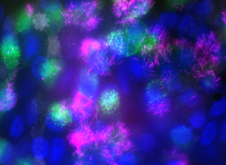
An indirect way of visualising viruses is to use antibodies (much like the ones your body makes in response to infection) to tag viruses with fluorescent molecules that give off light when they absorb certain types of radiation. We can even tag multiple things (such as virus and cellular components) with different colours so we can track more than one at the same time.
We can then detect the fluorescent light from the tags to see where viruses go inside our cells and what cell structures they interact with. This allows us to investigate things such as how drugs affect virus replication or how different strains of viruses behave differently.
Super resolution microscopy

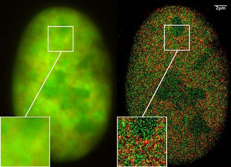
Recent advances in fluorescent microscopy have led to the development of super resolution microscopy, which combines very clever physics with computational methods to produce clear images that reveal highly detailed structures in cells.
Using this technique for virology can pinpoint areas of an infected cell with more accuracy. For instance, it can show exactly where inside the cell viruses are located, and what specific parts of cellular machinery viruses use to replicate.
Electron Microscopy

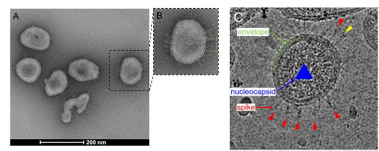
None of the techniques mentioned so far are able to directly visualise virus particles. That’s where electron microscopy comes in, as it can produce images at the nanometre scale. It does this by firing electrons at a sample and seeing how they interact with it. A computer then interprets this information to produce an image.
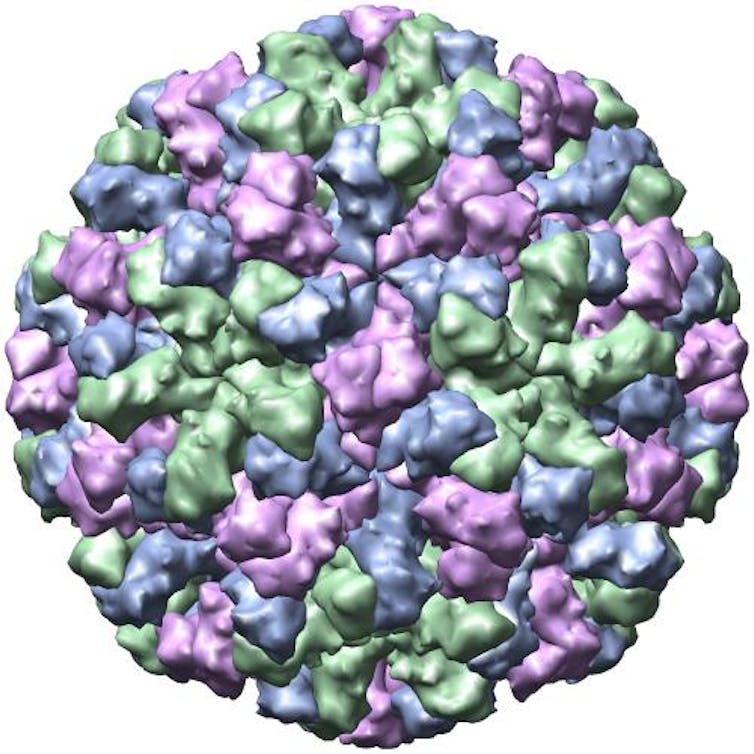
This enables us to visually investigate different stages of virus infection inside cells. Electron microscopy can also be used to visualise whole virus particles, as shown in the image above. From these images, we can form 3D structures of whole virus particles by computationally assembling images of thousands of particles taken in different orientations, such as this example of a 3D EM rendering of SARS-CoV-2.
Electron microscopy has been used for SARS-CoV-2 to determine how the virus uses its outer “spike” protein to interact with our cells and infect them. Such studies are really useful in working out how the virus gains access to our cells so we can work out how to use drugs to block it.
Evaluating the structure of the exterior of virus particles is also a great tool for identifying which antibodies can neutralise a virus, which can help produce more precise and effective vaccines.
Crystallography

Crystallography allows us to view structures in even more detail, at the atomic level. To do this, you need a really pure sample of virus (with no debris) suspended in solution. The liquid of the suspension is evaporated which causes crystallisation of the remaining solids (including the virus). These align in a uniform manner to form crystals that can then be exposed to X-rays.
A detector records the way in which the X-rays diffract (or “bounce”) from the crystallised sample, indicating where the electrons are in the sample structure. This information can then be used to construct an atomic-scale 3D structure of the sample.
As with electron microscopy, crystallography can be used to determine structures of viruses, such as the spike protein of SARS-CoV-2. Understanding these structures, especially how they interact with our cells and antibodies informs vaccine and drug design.
This article is republished from The Conversation under a Creative Commons license. Read the original article.

